图 3.
Typical case 3
典型病例 3
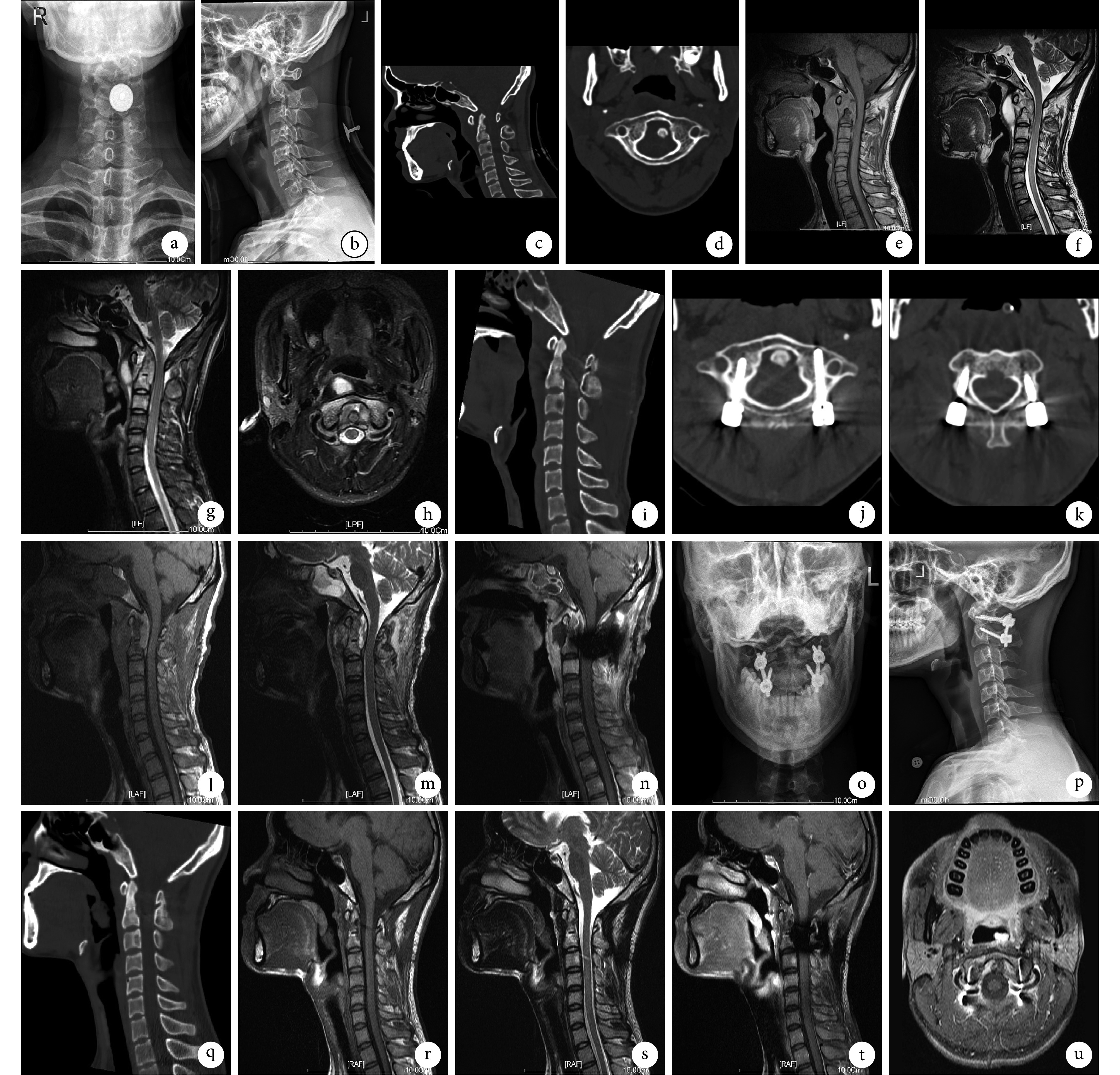
a、b. 术前正、侧位 X 线片;c、d. 术前矢状位、轴位 CT;e~h. 术前 T1、T2 及强化矢状位、轴位 MRI;i~k. 术后即刻矢状位、C1 及 C2 轴位钉道扫描 CT;l~n. 术后即刻 T1、T2 及强化矢状位 MRI;o~q. 术后 3 个月正、侧位 X 线片及矢状位 CT;r~u. 术后 6 个月 T1、T2 及强化矢状位、轴位 MRI
a, b. Preoperative anteroposterior and lateral X-ray films; c, d. Preoperative sagittal and axial CT; e-h. Preoperative T1, T2, enhanced sagittal, and axial MRI; i-k. CT of sagittal view and axial views of nail-tract scanning of C1 and C2 at immediate after operation; l-n. T1, T2, and enhanced sagittal MRI at immediate after operation; o-q. Anteroposterior and lateral X-ray films and sagittal CT at 3 months after operation; r-u. T1, T2, enhanced sagittal, and axial MRI at 6 months after operation

