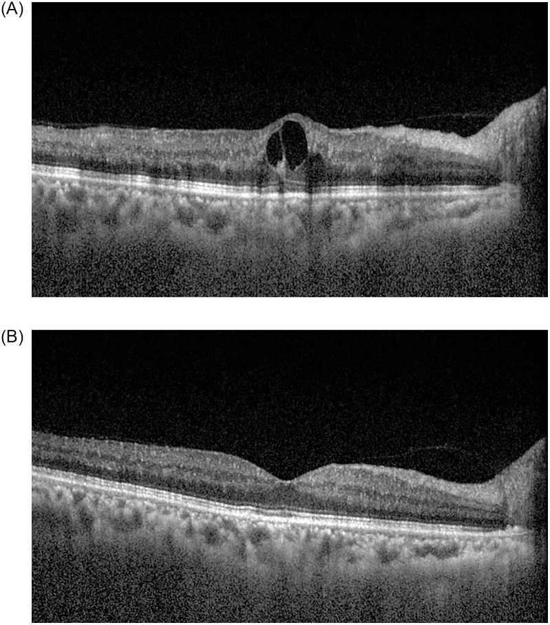
Fig 1. A 63-year-old female patient with central retinal vein occlusion in the left eye.
She had no obvious medical history. At the time of diagnosis, best-corrected visual acuity (BCVA) was 0.8 logMAR in the left eye, and diffuse flame-shaped retinal hemorrhages with macular edema was noted in the fundus. Central macular thickness (CMT) was 435.0 μm in the left eye (A). One month after the first intravitreal bevacizumab injection, BCVA in the right eye improved to 0.3 logMAR. The retinal hemorrhages improved, and CMT decreased to 296.0 μm in the left eye (B). The aqueous endothelin-1 level was 12.1 pg/mL at the time of the first intravitreal bevacizumab injection and decreased to 6.9 pg/mL one month after the first injection.

