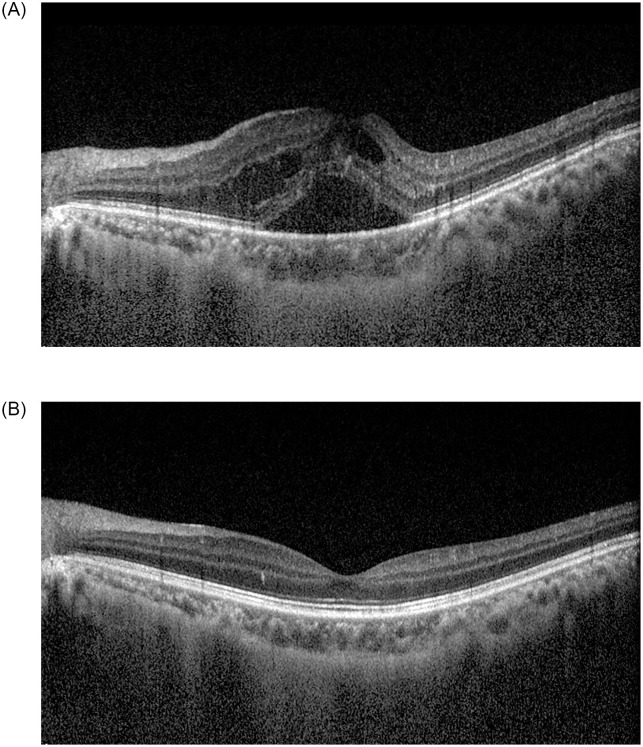
Fig 2. A 40-year-old male patient with branch retinal vein occlusion in the right eye.
He had a history of dyslipidemia. At the time of diagnosis, best-corrected visual acuity (BCVA) was 0.3 logMAR in the left eye. A fundus examination revealed flame-shaped retinal hemorrhage and cotton-wool spots along the superotemporal vascular arcade in the left eye. The central macular thickness (CMT) was 586.0 μm in the left eye (A). One month after intravitreal bevacizumab injection, BCVA in the left eye improved to 0.2 logMAR. The retinal hemorrhages improved, and CMT decreased to 286.0 μm in the left eye (B). The aqueous endothelin-1 level was 12.8 pg/mL at the time of the first intravitreal bevacizumab injection, and then decreased to 1.7 pg/mL one month after injection.

