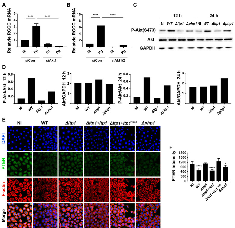
Fig 6. Control of RGCC by P. gingivalis involves Akt and PTEN.
TIGK cells were transiently transfected with siRNA to Akt1 (A) or Akt1/2 (B) or scrambled siRNA (siCon) and challenged with P. gingivalis (Pg). Expression of RGCC mRNA was measured by qRT-PCR. Data are expressed relative to noninfected (NI) controls. C) Western blots of TIGKs challenged P. gingivalis WT, Δltp1, Δphp1, or left uninfected (NI) for 12 h or 24 h. Blots were probed with antibodies to Akt, phospho(P-) Akt (serine 473) or GAPDH as a loading control. D) Densiometric analysis of blot in (C) using ImageJ. E) Fluorescent confocal microscopy of TIGK cells challenged with P. gingivalis WT, Δltp1, Δphp1, Δltp1+ltp1, Δltp1+ltp1C10S, or left uninfected (NI). Cells were probed with PTEN antibodies and Alexa Fluor 488 secondary antibody (green). Actin (red) was stained with Texas Red-phalloidin, and nuclei (blue) stained with DAPI. Cells were imaged at magnification ×63, and shown are projections of z-stacks generated with Volocity. F) PTEN staining intensity was quantified in over 200 cells and normalized to the number of cells with Volocity software. * p< 0.05, **** p < 0.001 compared to NI.

