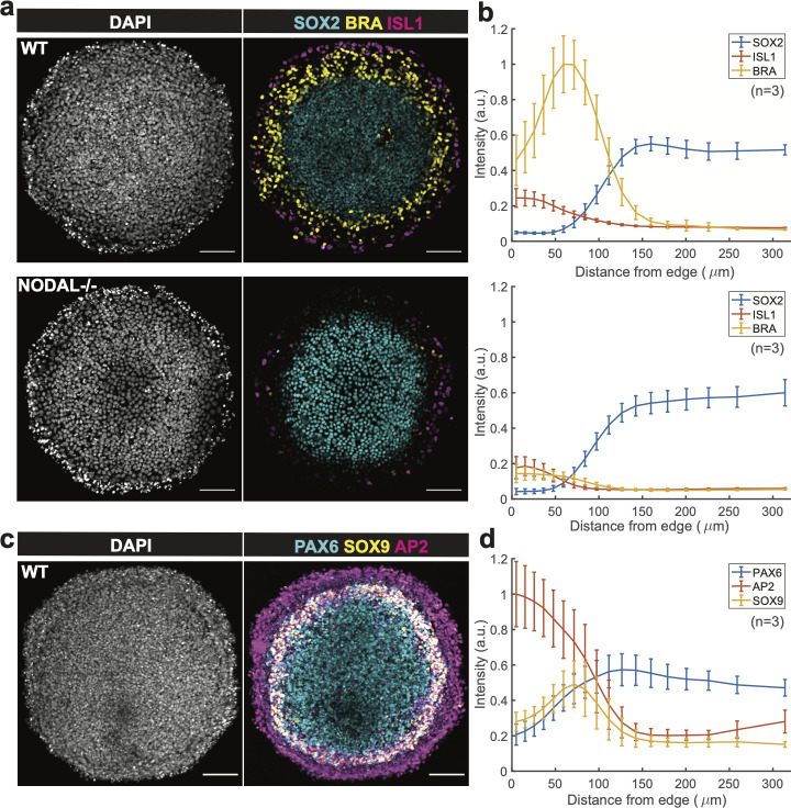
Fig 5. Reproduction of gastrulation and neural ectodermal induction results with the PEGDA-based micropatterning method.
(a) Confocal images of cell colonies immunostained for SOX2, BRA, and ISL1 at 48 h post BMP treatment in different conditions: Control and NODAL −/− cells were treated with 50 ng/ml BMP4. BMP, Bone Morphogenic Protein. Scale bar = 100 μm. Colony size: Diameter of 650 μm. (b) SOX2, BRA, and ISL1 levels of the wild type group (top) and Nodal -/- group (bottom) were quantified as a function of distance from the colony center (n = 3). (c) Confocal images of cell colony immunostained for PAX6, SOX9, and AP2. Patterns were initially treated for 3 days in N2B27 media with 10 ng/mL SB and then subsequently induced for 1 day in N2B27 media with SB, BMP4 and IWP2. Scale bar = 100 μm. Colony size: Diameter of 650 μm. (d) PAX6, SOX9, and AP2 levels of the ectodermally induced group were quantified as a function of distance from colony center (n = 3).

