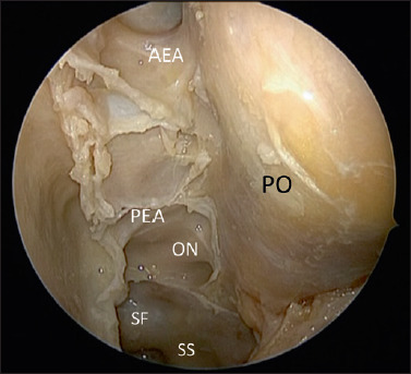
Figure 3.

Anatomic dissection showing the periorbit (PO) after complete spheno-ethmoidectomy with the exposure of the Skull base from the I fovea ethmoidalis, anterior ethmoidal artery (AEA), Posterior ethmoidal artery (PEA); to the sphenoid sinus and the following structures: Optic nerve (ON), Interoptic carotid recess and sellar floor Sphenoid Sinus (SS).
