Figure 4.

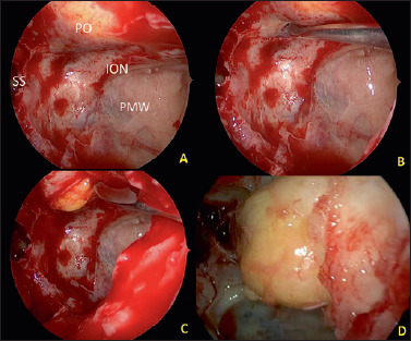
(A-D) Intraoperative view of left orbital decompression with the exposure of the sphenoid sinus (SS), Posterior maxillary wall (PMW), infraorbital nerve (ION), with incision of the inferior periorbit and the final fat ernjation.

(A-D) Intraoperative view of left orbital decompression with the exposure of the sphenoid sinus (SS), Posterior maxillary wall (PMW), infraorbital nerve (ION), with incision of the inferior periorbit and the final fat ernjation.