
Figure 4.
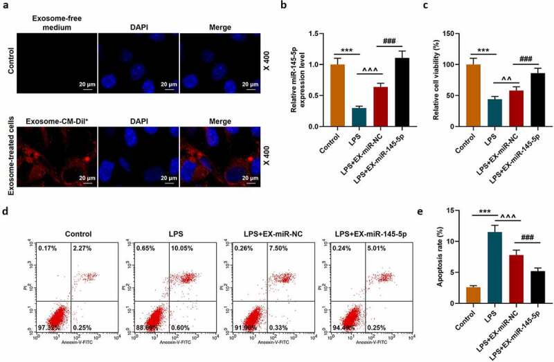
MSC-EXs could inhibit the effect of LPS inhibition ofon inhibiting miR-145-5p expression, cell viability and apoptosis in PC12 cells
(a) A cell injury model was constructed by treating PC12 cells with LPS. A fluorescence microscope was used to observe the changes in the uptake of fluorescently labeled EX by PC12 cells and the Control group. (b) QRT-PCR was used to detect the expression of miR-145-5p in PC12 cells. (c) MTT was used to detect the viability of PC12 cells treated with LPS. (d,e) Flow cytometry staining was used to detect cell apoptosis. (n = 3, ***P < 0.001, vs. Control group; ###P < 0.001, vs. LPS+EX-miR-NC group; ^^P < 0.01, ^^^P < 0.001, vs. LPS group)