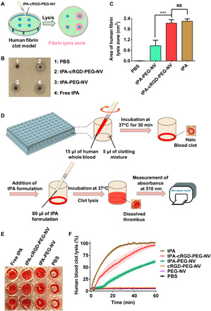
Fig. 5. Targeted fibrinolysis and thrombolysis under static conditions.

(A) Schematic illustration of the human fibrin clot model and the fibrin lysis zone induced by tPA-cRGD-PEG-NV. (B) Representative photograph of human fibrin clots and (C) calculated area of human fibrin lysis zone after treatment with PBS buffer (pH 7.4) only, tPA-PEG-NV, tPA-cRGD-PEG-NV and free tPA, respectively, which were preincubated with human activated platelets at 37°C for 2 hours. (D) Schematic illustration of the halo human blood clot assay protocol. (E) Representative photograph of human blood clots treated with PBS buffer (pH 7.4) only, tPA-PEG-NV, tPA-cRGD-PEG-NV, and free tPA, respectively, at 37°C for 1 hour. (F) Time-dependent clot lysis in the halo human blood clot model after treatment with PBS buffer (pH 7.4) only, PEG-NV, cRGD-PEG-NV, tPA-PEG-NV, tPA-cRGD-PEG-NV, and free tPA at 37°C for 1 hour, respectively. Data are presented as the average ± SD (n = 3). Statistical analysis was performed using the ANOVA (multiple comparisons) test. ***P < 0.001, and NS represents no significant difference between two groups.
