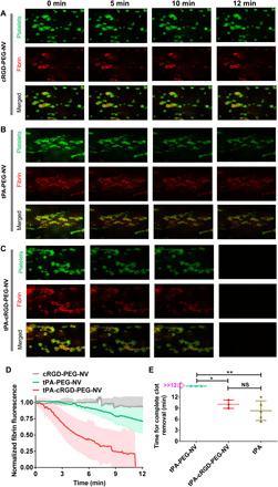
Fig. 7. Targeted thrombolysis under flow conditions.

Recalcified citrated blood labeled with DIOC6 and AF-647-FBG was perfused in the collagen- and TF-coated channels for 8 min at a shear rate of 1000 s−1. Channels were washed with HT buffer for 2 min, and then the recalcified blood containing (A) cRGD-PEG-NV, (B) tPA-PEG-NV, and (C) tPA-cRGD-PEG-NV, respectively, at an equivalent tPA concentration of 15 μg ml−1 was perfused in the thrombi-containing channels for the indicated time durations at a shear rate of 1000 s−1. The representative fluorescence images of platelets (green) and fibrin (red) at human thrombi were collected at different perfusion times. (D) Real-time changes in the red fluorescence of fibrin at human thrombi after perfusion with cRGD-PEG-NV, tPA-PEG-NV, and tPA-cRGD-PEG-NV, respectively. (E) Time required for complete human blood clot removal after perfusion with tPA-PEG-NV, tPA-cRGD-PEG-NV, and free tPA, respectively. Data are presented as the average ± SD (n ≥ 3). Statistical analysis was performed using the ANOVA (multiple comparisons) test. *P < 0.05 and **P < 0.01, respectively.
