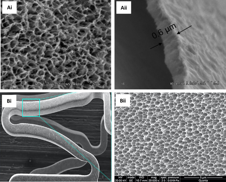
FIG. 7.
Representative SEM images of clinically tested porous stents. (a-i) Microporous hydroxyapatite-coated stent filled with sirolimus formulation (a-ii) cross section of the nanothin hydroxyapatite coating (∼600 nm). Reprinted with permission from Costa et al., JACC: Cardiovasc. Interventions 2(5), 422–427 (2008). Copyright 2008, Elsevier. (b-i) Nano+TM polymer-free stent showing strut microstructure after expansion. Strut thickness is approximately 91 μm having a large number of sirolimus-filled pores (∼400 nm) on the abluminal stent surface. (b-ii) Electron microscopy images of the nanopores (magnification ×20 000). Reprinted with permission from Liu et al., Catheterization Cardiovasc. Interventions 95(S1), 658–664 (2020). Copyright 2020 John Wiley and Sons.

