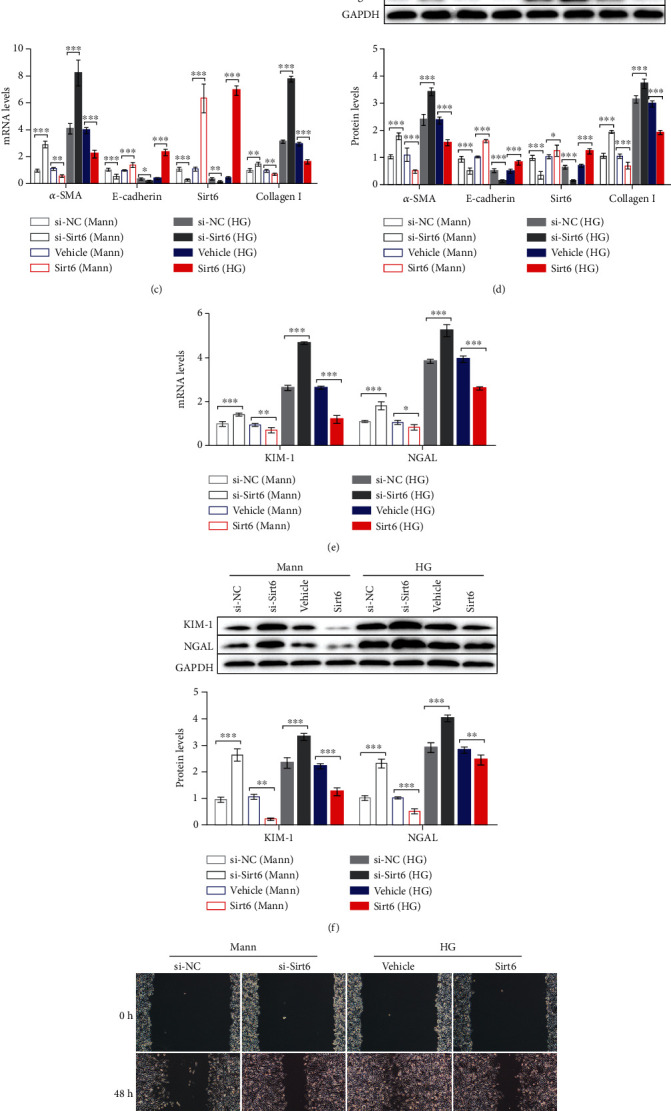
Figure 1.

The progression of EMT HG-induced HK2 cells is associated with reduced Sirt6 levels. (a, b) HK2 cells were cultured in the HG medium for 72 h. (a) mRNA expression of fibrotic genes and Sirt6 analyzed by qPCR. (b) Protein expression of fibrotic genes and Sirt6 assessed by western blot analysis. (c–f) HK2 cells were transfected with si-NC or si-Sirt6 to knock down Sirt6 or transfected with the vehicle or the Sirt6 overexpression vector to increase Sirt6 expression and cultured in the Mann medium or HG medium for 72 h. (c) mRNA expression of fibrotic genes and Sirt6 analyzed by qPCR. (d) Protein expression of fibrotic genes and Sirt6 assessed by western blot analysis. (e) mRNA expression of tubular damage genes analyzed by qPCR. (f) Protein expression of tubular damage genes assessed by western blot analysis. (g, h) Representative images of cell migration and graphs showing the wound area quantification in HK2 cells. After 48 h of transfection, wounds were generated using a 200 μl pipette tip and cultured for another 48 h. HK2 cells transfected with si-NC or si-Sirt6 were cultured in the Mann medium, and cells transfected with the vehicle or the Sirt6 overexpression vector were cultured in the HG medium. (i) Representative images of Transwell assay and graphs showing migrated cells. After 24 h of seeding, HK2 cells were transfected with si-NC or si-Sirt6 cultured in the Mann medium and transfected with the vehicle or the Sirt6 overexpression vector cultured in the HG medium. The data are presented as the means ± SD. n = 3 experiments in (a–i). ∗p < 0.05, ∗∗p < 0.01, and ∗∗∗p < 0.01.
