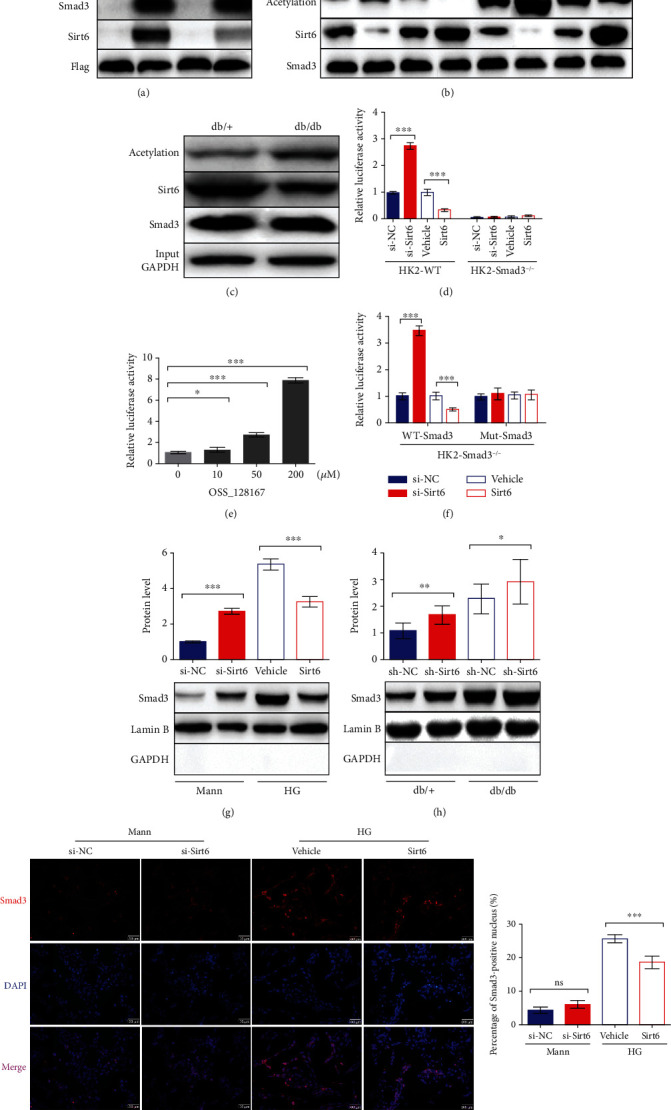
Figure 3.

Sirt6 inhibits Smad3 transcriptional activity by deacetylating it and suppressing its nuclear localization. (a, b) Exogenous and endogenous co-IP assays performed to assess the binding and between Sirt6 and Smad3 and Smad3 acetylation in HK2 cells. (a) HK2 cells cultured in the Mann or HG medium were transfected with Flag-Smad3 or Flag-IgG. Proteins immunoprecipitated with an anti-Flag antibody were analyzed using anti-acetylation, anti-Smad3, anti-Sirt6, and anti-Flag antibodies. (b) HK2 cells were cultured in the Mann or HG medium for 72 h. Proteins immunoprecipitated with an anti-Smad3 antibody were analyzed using anti-acetylation, anti-Sirt6, and anti-Smad3 antibodies. (c) Endogenous co-IP analysis to assess the binding and between Sirt6 and Smad3 and Smad3 acetylation in the kidneys of db/+ or db/db mice. Proteins immunoprecipitated with an anti-Smad3 antibody were analyzed using anti-acetylation, anti-Sirt6, and anti-Smad3 antibodies. Proteins from whole-cell lysates analyzed using anti-GAPDH antibodies. (d–f) The luciferase activity for the SBE-Luc reporter assay. (d) Wild-type HK2 cells and HK2-Smad3−/− cells cultured in the Mann medium were cotransfected with SBE-Luc plasmids with the si-NC, si-Sirt6, vehicle, and Sirt6 overexpression vector for 72 h, respectively. (e) Wild-type HK2 cells were transfected with SBE-Luc plasmids for 72 h. Different concentrations of OSS_128167 were added into the Mann medium to inhibit Sirt6 deacetylation at 24 h after transfection. (f) HK2-Smad3−/− cells stably overexpressing wild-type or mutant Smad3 with K333A and K378A mutations were medium-cotransfected with SBE-Luc plasmids with the si-NC, si-Sirt6, vehicle, and Sirt6 overexpression vector, respectively, and cultured in the Mann medium for 72 h. (g, h) Nuclear proteins were extracted from HK2 cells or the kidneys of db/db and db/+ mice. (g) Smad3 protein expression in HK2 cells transfected with the si-NC, si-Sirt6, vehicle, and Sirt6 overexpression plasmid, as assessed by western blot analysis. (h) Smad3 protein expression in the kidneys of db/db or db/+ mice injected with sh-NC or sh-Sirt6 lentivirus, as assessed by western blot analysis. (i) Representative IF images of HK2 cells staining with Smad3 and DAPI in different conditions. The data are presented as the means ± SD. n = 3 experiments in (a–g) and (i). n = 6 experiments in (h). ∗p < 0.05, ∗∗p < 0.01, and ∗∗∗p < 0.01.
