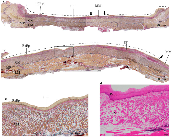
Fig. 2.
Representative histological findings of the artificial ulcer in esophagectomy specimen after ESD in the TA group. a Histological findings of the esophagectomy specimen from one representative case. The black arrow indicates the border of the ESD scar because of the muscularis mucosae disruption. The dotted line indicates the ESD ulcer scar. The subepithelial fibrous tissue is relatively thin over the whole area and the regenerated epithelium and the muscularis propria are close to each other. EVG, original magnification × 20. b Histological findings of the central part of an artificial ulcer in an esophagectomy specimen from another case. The black arrow indicates the disruption of muscularis mucosae, the dotted line indicates the ESD ulcer scar. The regenerated epithelium and the muscularis propria layer are very close, and subepithelial fibrous tissue between them is barely observed. EVG, original magnification × 20. c Enlarged view of the area surrounded by black squares in Fig. 1b. The subepithelial fibrous tissue is very thin and the regenerated epithelium and the muscularis propria layer are almost in contact with each other. EVG, original magnification × 40. d Histological findings of another case. The regenerated epithelium and the muscularis propria layer are almost in contact with each other. HE, original magnification × 40. ReEp regenerated epithelium, MM muscularis mucosae, MP muscularis propria, CM circular muscle, LM longitudinal muscle, SF subepithelial fibrous tissue, EVG Elastica van Gieson staining, HE hematoxylin and eosin staining, TA triamcinolone acetonide

