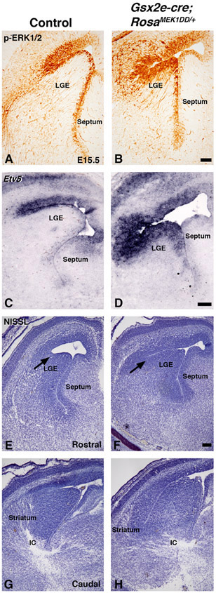
Figure 3: Sustained MEK/MAPK activity in the LGE of Gsx2e-Cre;RosaMEK1DD/+ embryos.
Representative images at E15.5 from controls (A, C, E, G) and double transgenic Gsx2e-Cre;RosaMEK1DD/+ (called MEK/MAPK GOF) embryos (B, D, F, and H). MEK/MAPK GOF embryos show increased p-ERK1/2 (B) and Etv5 (D) staining in the LGE ventricular zone compared to controls (A,C). NISSL staining shows abnormal morphology at the LGE sulcus (arrows in E-F), LGE, striatum, and forming axon tracts in the ventral telencephalon of MEK/MAPK GOF embryos (F,H) compared to controls (E,G). Scale bars in: B= 100μM for images in A-B, D= 200μM for images in C-D, F=200μM for images E-H. LGE= lateral ganglionic eminence, IC=internal capsule

