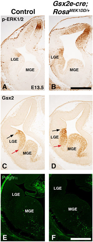
Figure 6: Sustained MEK/MAPK activity alters LGE morphology at early stages.
Representative images at E13.5 from controls (A, C, and E) and MEK/MAPK GOF double transgenic Gsx2e-Cre;RosaMEK1DD/+ (B, D, and F) embryos. p-ERK1/2 expression is increased at E13.5 in MEK/MAPK GOF embryos (B) compared to controls (A). Gsx2 is expressed in presumptive LGE area of MEK/MAPK GOF embryos but the high dorsal to low ventral gradient observed in controls (arrows in C) is more uniform in GOF embryos (arrows in D). Unlike later stages, MEK/MAPK GOF embryos at E13.5 do not show precocious Pdgfrα staining in the LGE. The Pdgfrα staining pattern in the ventral MGE of MEK/MAPK GOF embryos (F) is similar to controls (E). Scale bars in: B= 500μM for images in A-D. F= 500μM for images in E-F.

