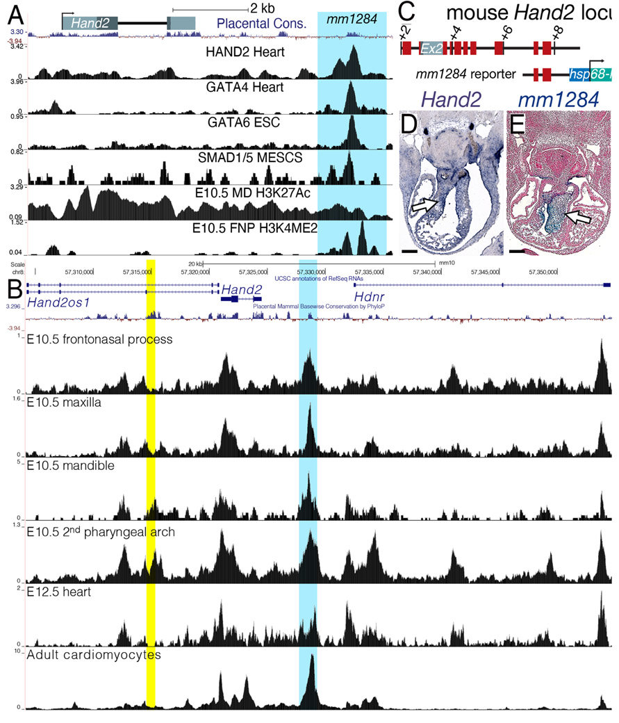
Figure 6. Identification of an endocardial cushion-specific Hand2 enhancer.
A) Schematic of the mouse Hand2 locus. Peaks of enrichment for HAND2, GATA4/6, and SMAD1/5 overlap with a region, evolutionarily conserved among placentals, that displays histone marks for active transcription in NCCs (H3K4ME2 in the E10.5 fronto-nasal processes (FNP) and H3K27Ac in the mandibular pharyngeal arch (MD). This mm1284 sequence is highlighted in blue. B) ATAC-seq from E10.5 frontonasal process, maxilla, mandible, and 2nd pharyngeal arch (Minoux et al., 2017), E12.5 hearts (Zhou et al., 2017), and adult cardiomyocytes (Monroe et al., 2019) (Yellow shading highlights the Hand2 mandibular arch enhancer. (Charite et al., 2001) C) Schematic of the mm1284+hsp68-lacZ reporter construct. Red blocks denote regions of significant sequence conservation D) Hand2 section in situ hybridization showing expression in the E11.5 OFT cushions (white arrow). E) Section of an X-gal stained E11.5 mm1284+hsp68-lacZ transgenic embryo showing expression in the OFT cushions (white arrow). n = 5/7. Scale bars = 250 μm.

