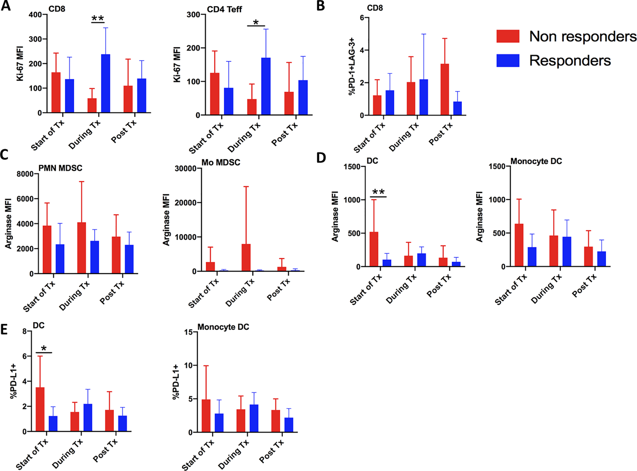
Figure 2. Improved peripheral anti-tumor immunity signature in responding versus non-responding patients.

Peripheral blood mononuclear cells were isolated from patients prior to, during, and post treatment, and assessed for immune composition and function by 20-color flow cytometry. A) Proliferation of proliferating CD8 and CD4 T effector cells was analyzed by Ki-67 expression B) Percentage of exhausted PD-1+ LAG-3+ CD8 T cells. C,D) Arginase expression was measured in C) immune suppressive PMN and Mo myeloid-derived suppressor cells, and in D) dendritic cells and monocyte-derived dendritic cells. E) Percent of dendritic cells and monocyte-derived dendritic cells expressing PD-L1 was also measured. ns, not significant; *, P < 0.05; **, P < 0.01; ***, P < 0.001; ****, P < 0.0001 by two-way ANOVA (Sidak’s multiple comparisons test)
