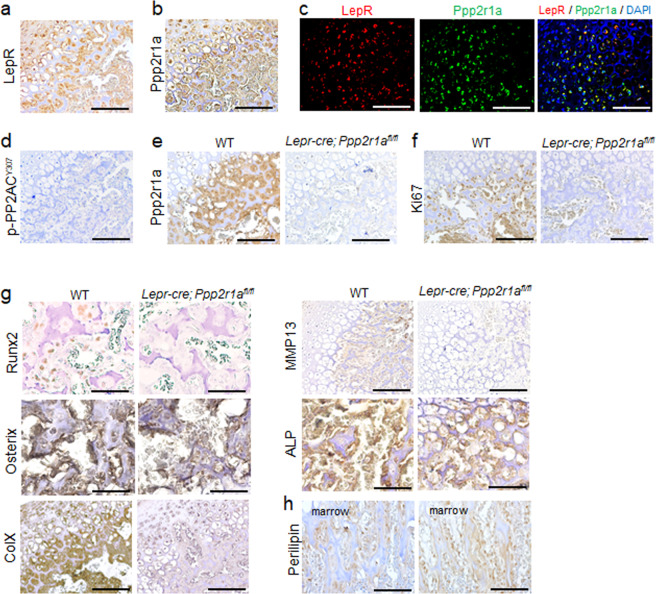
Fig. 4. Deletion of PP2A in LepR+ MSCs inhibited proliferation and hypertrophic differentiation of SOC in early postnatal stage.
Sections of P15 distal femur from Lepr-cre; Ppp2r1afl/fl mice were subjected to immunostaining. a–d Immunohistochemistry (IHC) and immunofluorescence reveal LepR+ MSCs express Ppp2r1a and unphosphorylated (Y307) PP2AC in SOC. (a) LepR IHC. b Ppp2r1a IHC. c Double immunofluorescence of LepR and Ppp2r1a. d Unphosphorylated (Y307) PP2AC IHC. e IHC reveals successful deletion of Ppp2r1a in LepR+ MSCs in SOC. IHC reveals that deletion of Ppp2r1a in LepR+ MSCs leads to decreased expression of Ki67 (f) and hypertrophic markers, such as Runx2, Osterix, collagen X, MMP13 and alkaline phosphatase (g), and increased expression of Perilipin (h) in SOC at P15. HZ = Hypertrophic zone; POC = Primary ossification center; ColX =collagen X; ALP = alkaline phosphatase. Scale bar, 100 μm.

