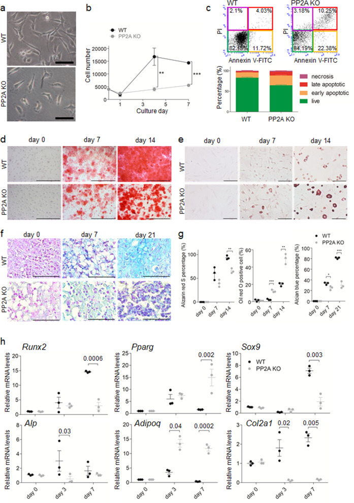
Fig. 6. PP2A deletion in LepR+ MSCs inhibits osteogenesis and chondrogenesis but promotes adipogenesis in vitro.
MSCs isolated from WT (WT MSCs) and Lepr-cre; Ppp2r1a fl/fl mice (PP2A KO MSCs) were subjected to analysis. a WT and PP2A KO MSCs exhibit no difference in morphology. Scale bar, 50 μm. b PP2A KO MSCs exhibit decreased proliferation. n = 3 independent experiments. c Represented flow cytometry plots of Annexin V-FITC/PI assay for apoptosis analysis of WT and PP2A KO MSCs. After FSC/SSC gating (left), MSCs are categorized into necrosis, late apoptosis, early apoptosis and live stages (right). The corresponding percentage are shown in bar chart. d WT and PP2A KO MSCs were induced for osteogenic differentiation and stained with Alizarin Red S at different time points. Scale bar, 1000 μm. e WT and PP2A KO MSCs were induced for adipogenic differentiation and stained with Oil red O at different time points. f WT and PP2A KO MSCs in micromass were induced for chondrogenic differentiation and stained with Alcian blue & nuclear fast red at different time points. Scale bar in e, f, 100 μm. g Quantification percentage of Alizarin red S staining, Oil red O staining and Alicante blue staining according to different differentiations respectively at each time points. n = 3 independent experiments. h qPCR analysis of transcript levels for genes associated with osteogenesis (Runx2, Alp), adipogenesis (Pparg, Adipoq) and chondrogenesis (Sox9, Col2a1) differentiation in MSCs induced by differentiation medium at each time points. Transcript levels were normalized based on β-actin amplification. n = 3 independent experiments. *p < 0.05; **p < 0.01; ***p < 0.001 as determined with Student’s t-test. Data are mean ± s.d.

