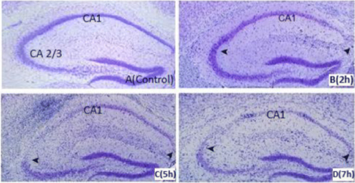
Fig. 8.
Nissl staining in the hippocampus of the Control (a) and 2, 5, and 7 h/days exposure to DEPs for 5 days/week within 12 weeks. By 5 h/d exposure, neurons of CA1and CA3 region are lost in the mouse (c) but minimal neuronal dropout is evident in the 3 h/day exposure mouse (b). By 7 h/d exposure, the majority of CA1 and CA3 neurons are lost in mice (d). Hippocampal CA1 and CA3 neurons were loose and absent; nucleoli were missing or indistinct (arrows). In the control group, neurons were fairly well preserved and sparse in the CA1 and CA3 sub-region. Sections are representative of at least 4 mice of each group

