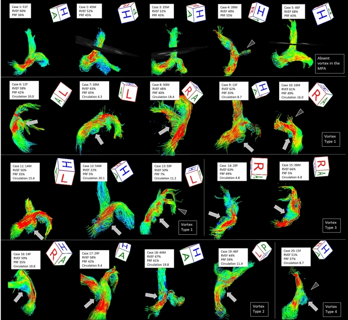
Figure 5.
Streamline imaging flows of the pulmonary arteries of 20 patients with repaired Tetralogy of Fallot. Case 1–5 did not show a vortex in the main pulmonary arteries (MPA), however, there were vortical or helical flows in the right and left PA in some patients. Case 6–20 showed vortex formation in the MPA. Vortices typically originated posteriorly in the MPA (Type 1: Case 6–13) and formed helices. Other vortices originated from the MPA bifurcation to the right PA (Type 2: Case 16–19), at the proximal right PA (Type 3: Case 14, 15), or from the MPA bifurcation to the left PA (Type 4: Case 20). Arrows indicate vortices and arrowheads indicate the left pulmonary artery stenosis. F female, M male, RVEF right ventricular ejection fraction, PRF pulmonary regurgitant fraction, Circulation longitudinal integral circulation value. Flow visualization was performed using 4D flow postprocessor software (iT Flow; Cardioflow Design. Inc., Tokyo, Japan).

