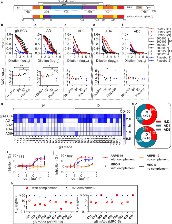
Fig. 4. Complement-enhanced neutralizing activity of gB antibodies.
a Schematic representation of the full-length HCMV gB (top, #ACL51135) and gB-ectodomain (gB-ECD) (bottom). Disulfide bonds are represented as black brackets, antigenic domains (AD1, AD2, AD4, AD5) are indicated in black brackets. Structural domains are labeled as, signal sequence (SS), membrane-proximal region (MPR), transmembrane domain (TM), cytoplasmic domain (Cyto). Numbers denote construct boundaries. b–f ELISA measuring plasma antibody binding to gB-ECD (b), AD1 (c), AD2 (d), AD4 (e), and AD5 (f). The top panel shows optical density units at OD450 nm (Y axis) and reciprocal plasma dilutions (X axis). The sera of HCMV-seropositive subjects (HCMV+), the placebo control (Placebo), and the vaccinated subjects are indicated. The bottom panel shows the normalized area under the curve (AUC) of the ELISA. An unpaired two-tailed t test was performed. Differences with statistical significance are shown with p values, “**” indicates p < 0.001. “*” indicates p < 0.05. The data are representative of two independent experiments. g Thirty-seven gB mAbs recognize four antigenic domains as determined by ELISA. The recombinant gB-ECD, AD1, AD2, AD4, and AD5 were coated at 3 µg/ml on ELISA plates, and tested for reactivity with gB antibodies. The heatmap indicates the value of OD450 nm of the mAbs to different gB domains in ELISA. h Distribution and binding specificity of gB mAbs from the intramuscular injection group (IM) and the intradermal injection group (ID). i, j The gB mAb (175 and 892) was mixed with AD169r-GFP virus with or without rabbit complement in neutralizing assay. The neutralizing activity is detected by GFP+ cell counting at 44 hours after infection. Error bars indicate the standard deviation. k Complement-enhanced neutralizing activity of nine gB antibodies in ARPE-19 and MRC-5 cells. The IC50 values defined as antibody concentrations required to neutralize 50% of viral infectivity were calculated by four-parameter curve fitting. The rabbit complement was added at 1:64 final dilution. The results shown are representative of two independent experiments.

