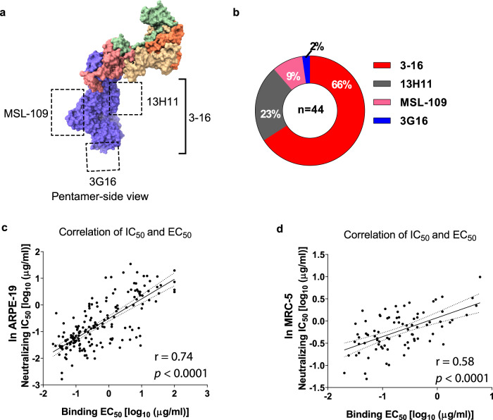
Fig. 5. The gHgL mAbs bind to diverse epitopes on the gHgL.
a The binding sites of four gHgL reference antibodies: 3–16 binds to the gH of pentamer, 13H11 binds to gHgL kinked region, 3G16 binds to the C-terminal domain of gH, MSL-109 binds to the H2 domain of gH as indicated. b Forty-four gHgL mAbs bind to overlapping regions with the four reference mAbs: 66% to the 3–16 epitope, 23% to the 13H11 epitope, 9% to MSL-109 epitope, and 2% to the 3G6 epitope. c, d Neutralization potency (IC50) of the gHgL mAbs in ARPE-19 cells (c), and MRC-5 cells (d), positively correlates with their binding affinity (EC50) to the HCMV virion. The IC50 and EC50 values shown are representative of two independent experiments. The linear association was calculated by the Pearson correlation, two-tailed. The Molecular graphic images of pentamer (PDB: 5VOD) were produced using UCSF ChimeraX (http://www.cgl.ucsf.edu/chimerax/).

