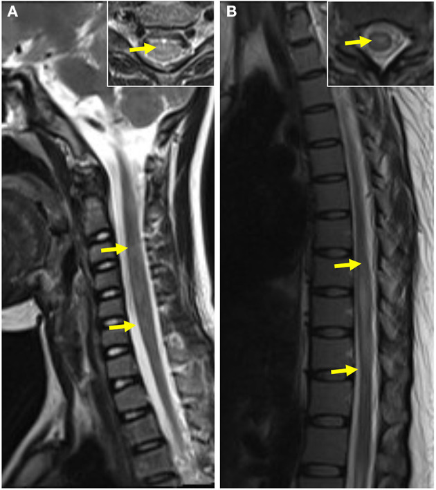
Figure 5.

Spinal cord MRI of pediatric and adult patients. (A) T2-weighted MRI of the spinal cord of a pediatric patient showed abnormal patchy signals at the cervical segment The coronal image of lesion-involved slices was showed in the right box. (B) T2-weighted MRI of the spinal cord of an adult patient showed a striped hyperintense lesion at the thoracic segment. The coronal image of lesion-involved slices was showed in the right box.
