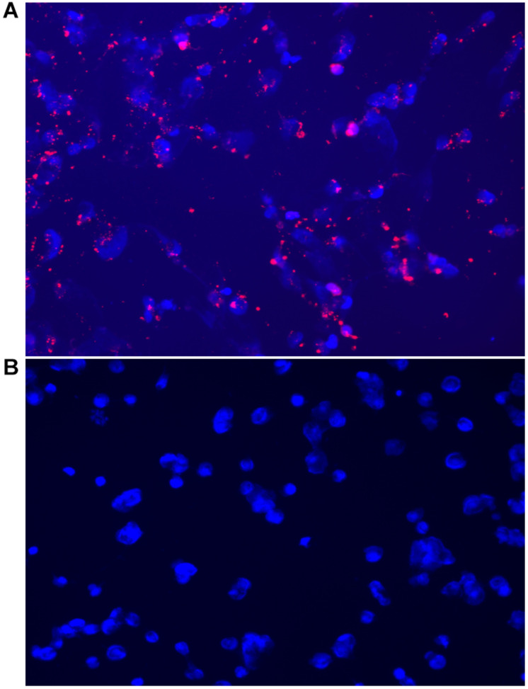
FIGURE 6.
Fluorescent microscopy images acquired on the CELENA® X High Content Imaging System of (A) OVCAR-5 cells incubated with MSN_CurNQ for 24 h (20× magnification). Blue fluorescence is DAPI nuclear staining and red fluorescence is MSN_CurNQ. (B) Cells given no treatment (control). No red fluorescence is present in the control group, confirming fluorescence is from MSN_CurNQ.

