. 2021 Mar 9;13(2):223–228. doi: 10.4055/cios20097

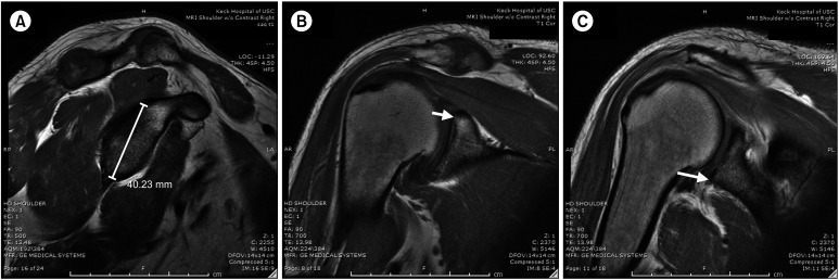
Fig. 2. (A) Sagittal T1-weighted magnetic resonance imaging (MRI) with glenoid height. The ruler head was measured from the superior to inferior articular surfaces of the glenoid on the coronal image for confirmation. (B) Coronal T1-weighted MRI with superior glenoid labelled. The superior glenoid is indicated by the white arrow. Using the crosshatch localization feature, confirmation of the glenoid height was attained on the sagittal image. (C) Coronal T1-weighted MRI with inferior glenoid labelled. The inferior glenoid is indicated by the white arrow. Using the crosshatch localization feature, confirmation of the glenoid height was attained on the sagittal image.

Fig. 2