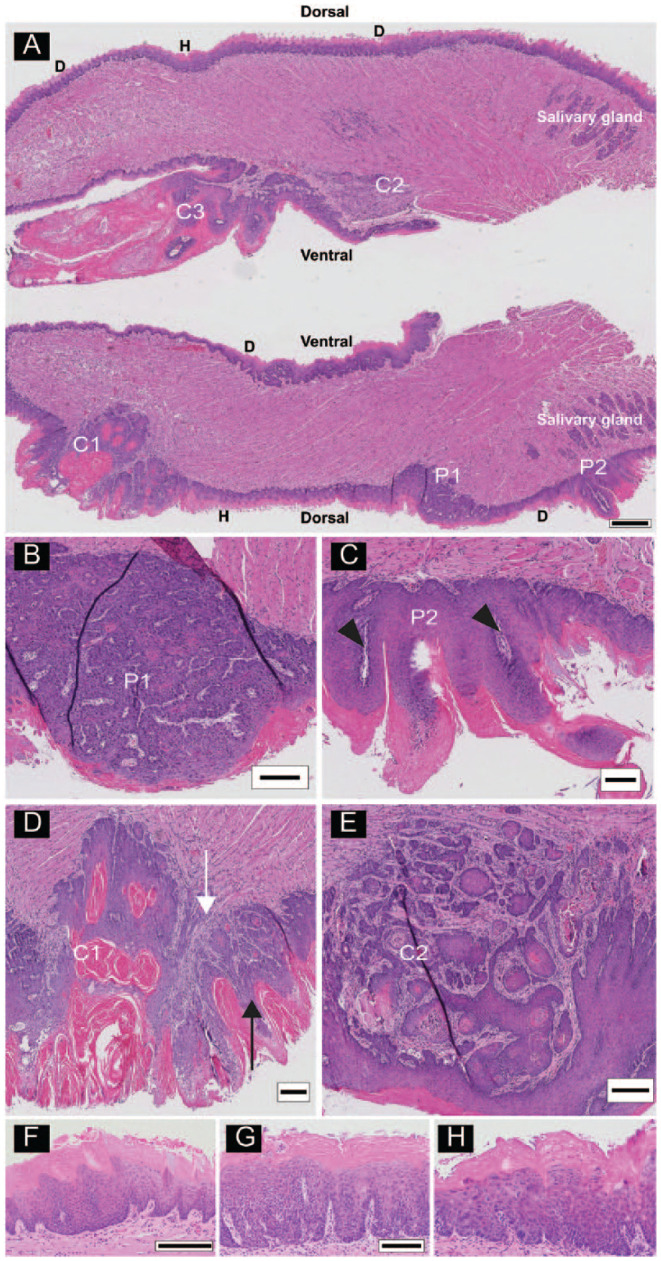
Figure 1.

Multiple lesions on a tongue from a 4-nitroquinoline 1-oxide (4NQO)–treated animal. (A) Longitudinal sections (anterior to the left) of a bisected tongue. Tongues were sectioned from the center outward. The scanned images are from slide 40, so that the top and bottom portions of the tongue are separated by 390 µm. Lesions include acanthosis, hyperkeratosis (H), field dysplastic changes (D), papillomas (P1, P2), and cancers (C1, C2, C3). (B) High-power image of P1, a dorsal sessile papilloma. (C) High-power image of P2, a dorsal pedunculated papilloma with finger-like epithelial proliferations with fibrovascular cores (arrowheads). (D) High-power image of C1, a dorsal papillary squamous cell carcinoma (pSCC) with exophytic (black arrow) and invasive (white arrow) components. (E) High-power image of C2, a ventral invasive squamous cell carcinoma (iSCC). High-power images of additional lesion types from other tongue sections. (F) Acanthosis and hyperkeratosis. (G) Hyperkeratosis with low-grade dysplasia and (H) high-grade dysplasia. Scale bars: A = 500 µm, B–E = 250 µm, F = 200 µm, G and H shown in G = 100 µm.
