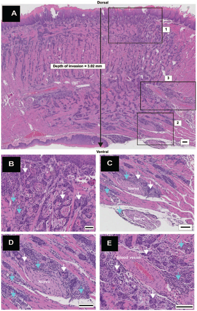
Figure 2.

Deeply invasive squamous cell carcinoma (diSCC) with aggressive features. (A) Longitudinal section (anterior to the left) shows a diSCC involving the entire dorsoventral thickness of the tongue with depth of invasion greater than 2 mm. High-power views of inset boxes 1, 2, and 3 shown in panels B–D. (B) Inset box 1: islands of diSCC (white arrows) invading stroma with interspersed inflammatory cells (turquoise arrows). (C) Inset box 2: Perineural invasion (PNI) with cancer (white arrows) surrounding greater than 50% of a nerve at multiple foci. Inflammation (turquoise arrows) is interspersed between nerves and cancer. (D) High-power image of PNI, cancer (white arrows), and inflammation (turquoise arrows). (E) Inset box 3: large blood vessels surrounded by cancer islands (white arrows) and inflammation (turquoise arrows). Scale bars: A = 100 µm; B–E = 50 µm.
