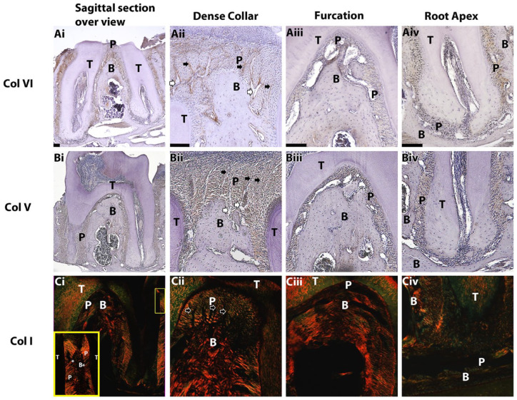
Figure 4.
Immunohistochemistry, sagittal slices. Brown staining for collagen VI (Ai–Aiv) and V (Bi–Biv) represents higher protein concentration. Polarized light and picrosirius red staining were used for collagen I (Ci–Civ); yellow represents higher collagen density and alignment along the sectioning plain, whereas green stain is indicative of orientation perpendicular to the sectioning plain. Yellow frame in Ci: inset of yellow rectangle in top right. Black arrows point to longitudinal structures in the dense collar, and white arrows show interstitial spaces forming next to alveolar bone. B, alveolar bone; P, periodontal ligament; T, tooth. *Abrupt change of periodontal ligament dense collar fibers direction. Scale bars: 100 µm.

