Figure 2.

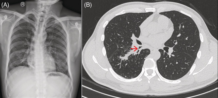
Chest X‐ray and chest computed tomography (CT) scan. (A) Chest X‐ray showed no abnormal finding of lung fields. (B) Chest CT scan showed the narrowing of the right lower lobe bronchus (red arrow).

Chest X‐ray and chest computed tomography (CT) scan. (A) Chest X‐ray showed no abnormal finding of lung fields. (B) Chest CT scan showed the narrowing of the right lower lobe bronchus (red arrow).