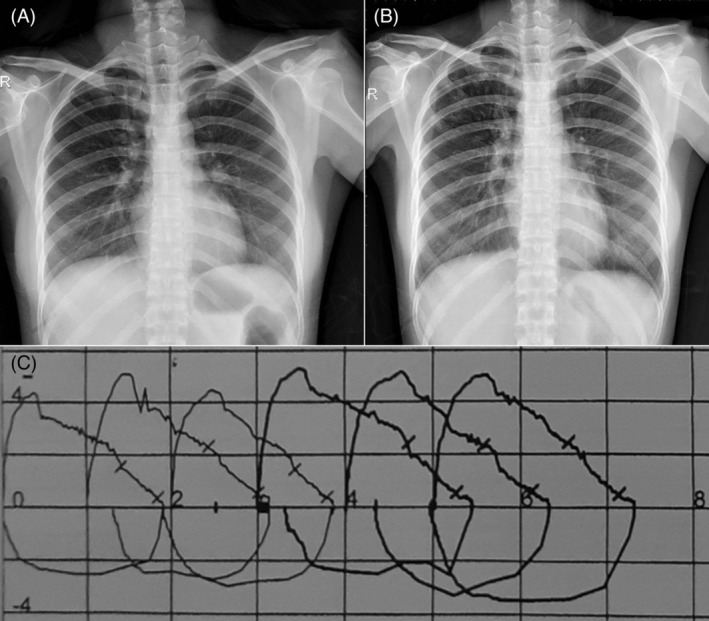
Figure 3.

Two chest X‐rays and the flow–volume curve of spirometry. (A) Chest X‐ray (CXR) at the first visit revealed the right apical fibrosis. (B) Second CXR after more than four months of the first one revealed the right apical infiltration. (C) The flow–volume curve showed a positive bronchodilator test with bold curve representing post‐test spirometry.
