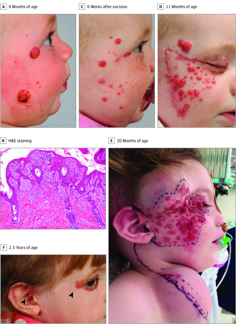
Figure 1. Clinical Progression of Agminated Spitz Nevi.
A, At presentation, 2 large exophytic, erythematous, hemorrhagic nodules affected the right cheek, and smaller erythematous papules were observed on the right temple, cheek, and upper eyelid. B, Hematoxylin-eosin (H&E) staining revealed a polypoid, symmetrical lesion comprising intradermal and junctional clusters of plump epithelioid and spindle nevus cells with occasional Kamino bodies. C, Six weeks after excision of the 2 larger lesions, the smaller erythematous papules enlarged substantially, and more lesions appeared on the right cheek and temple. D, By 11 months of age and despite multiple further excisions, more lesions continued to arise in a segmental distribution involving the right cheek, temple, and the upper and lower eyelids. E, Preoperative appearance at age 20 months showing extensive lesions involving the right cheek, with tissue expanders in situ at the right temple and right neck placed previously to facilitate reconstructive surgery. F, The patient at 2.5 years of age after resection of most of the affected tissue from the right cheek and temple and cervicofacial flap reconstruction. Residual periocular and auricular lesions (arrows) had increased in size.

