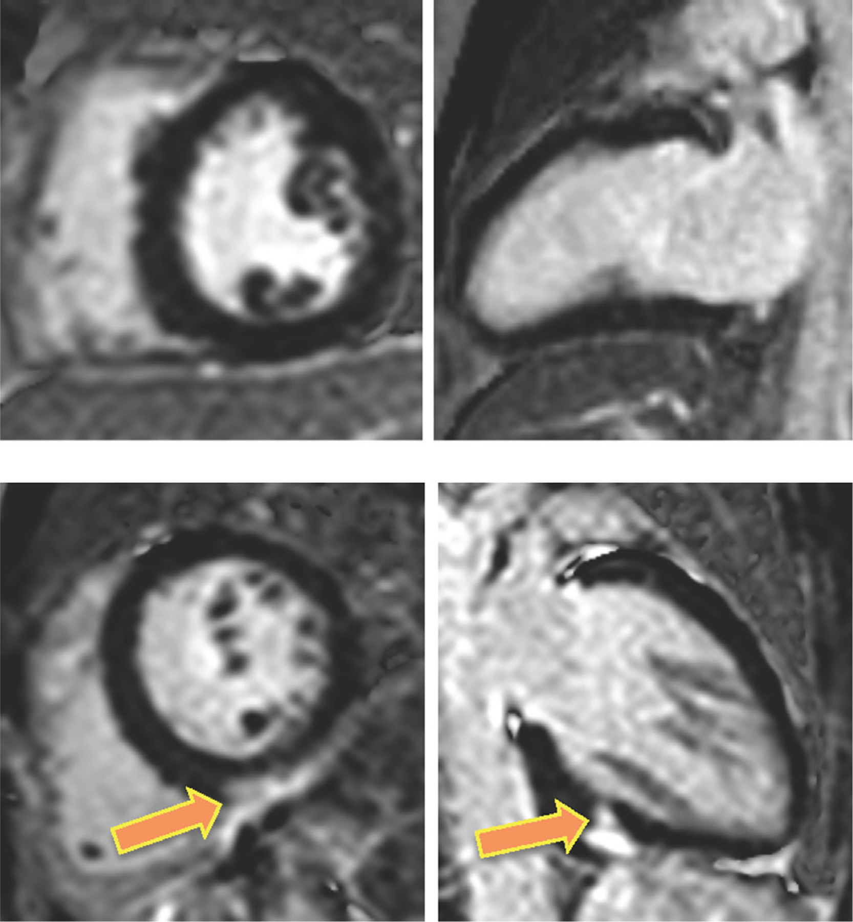
Figure 1.

CMR images obtained in two patients with extra-cardiac sarcoidosis and RV dysfunction: one without cardiac involvement (top) and the other with LGE in the LV myocardium (bottom), indicative of CS. Note atypical mid-inferior pattern of LGE in this latter patient (arrows).
