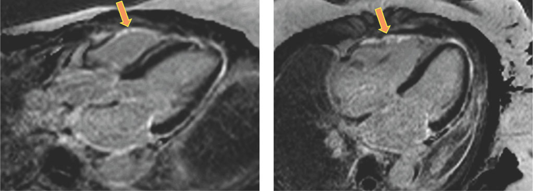
Figure 4.

CMR images obtained in a patient with extra-cardiac sarcoidosis and RV dysfunction, without LGE in the left ventricular myocardium, who had LGE in the right ventricular free wall (arrows). Of note, this finding could not be used to diagnose RV involvement with a similar level of confidence in other study patients.
