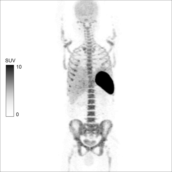
Figure 1.
MIP image of 18F-FDG-labelled WBC whole body PET/CT in a patient with aortic graft infection following antibiotic therapy. The image shows physiological distribution in the spleen, liver and the bone marrow. 11 The labelling efficiency of the radiopharmaceutical in this case was 90.3%. No area of abnormal tracer uptake is seen. However, mild brain uptake due to minimal free 18F-FDG activity and excretion via the kidneys into the urinary bladder is also noticed. FDG, fluoro-D-glucose; MIP, maximum intensity projection; PET, positron emission tomography; SUV, standardized uptake value; WBC, white blood cell.

