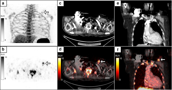
Figure 4.
A 72-year-old male with dilated cardiomyopathy had a past history of pacemaker implantation in the left chest wall. He then developed pus discharge from the pacemaker site, and hence the pacemaker was removed and reimplanted on the right side. He still had pus discharge from the left chest wall with a negative pus culture. MIP image (a) of the regional 18F-FDG-labelled WBC PET/CT performed with a suspicion of persistent infection showed a focus of tracer uptake in the left chest wall (B, white arrow, SUVmax 8.9) which localized to the pacemaker leads in the left chest wall on the axial (c, d) and coronal (e, f) CT and fused PET/CT images. A repeat pus culture from the tracer-avid site revealed infection with Citrobacter sedlakii. Subsequently the pacemaker leads were removed and the patient became asymptomatic. FDG, fluoro-D-glucose; MIP, maximum intensity projection; PET, positron emission tomography; SUV, standardized uptake value; WBC, white blood cell.

