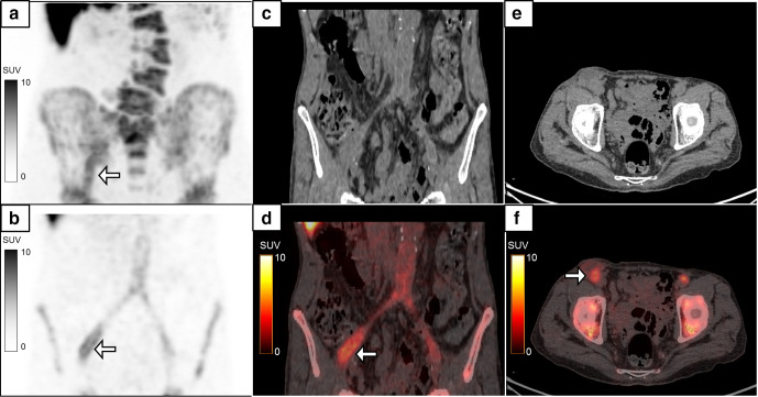
Figure 5.
A 74-year-old male, a case of aortoiliac occlusive disease, underwent aorto-bifemoral bypass and prosthetic grafting 1 year back. He presented with complaints of pus discharge from the wound in the right groin, which was positive for infection with Proteus mirabilis. MIP image (a) of the 18F-FDG-labelled WBC PET/CT done to assess extent of infection showed linear tracer activity in the right iliac region which on the coronal images (b–d, white arrows, SUVmax 5.2) localized to the collection along the bypass graft in the right hemipelvis extending to the right groin (e, f, white arrow). The patient recovered following debridement and drainage of the pus along with a course of intravenous antibiotics. FDG, fluoro-D-glucose; MIP, maximum intensity projection; PET, positron emission tomography; SUV, standardized uptake value; WBC, white blood cell.

