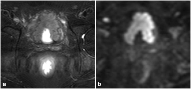
Figure 2.
60-year-old male with high-grade, muscle invasive urothelial cancer. (A) Axial T 2 weighted images showing an irregular plaque-like mass arising from the anterior and both lateral walls of the urinary bladder. The bladder is poorly distended secondary to the tethering effect of the tumor on the bladder wall. (B) Axial DWI b1500 images showing marked diffusion restriction of the tumor. DWI, diffusion-weighted imaging.

