Figure 1.
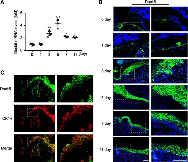
Expression pattern of Dock5 in wounds. Full-thickness wounds were made on the dorsum of C57BL/6 mice. A: mRNA expression of Dock5 in wound biopsies at the indicated time points after injury was analyzed by quantitative real-time PCR. B: In situ hybridization was performed using a Dock5-specific probe. Green indicates Dock5 expression, and blue indicates DAPI. C: Wound sections from C57BL/6 mice were stained for Dock5 (green) and cytokeratin 14 (CK14) (red, keratinocyte marker). Dotted lines in B and C indicate magnified area. Scale bars, 100 μm for B and C. n = 3 mice per group for A–C. Data are presented as means ± SEMs. *P < 0.05, **P < 0.01, ***P < 0.001.

