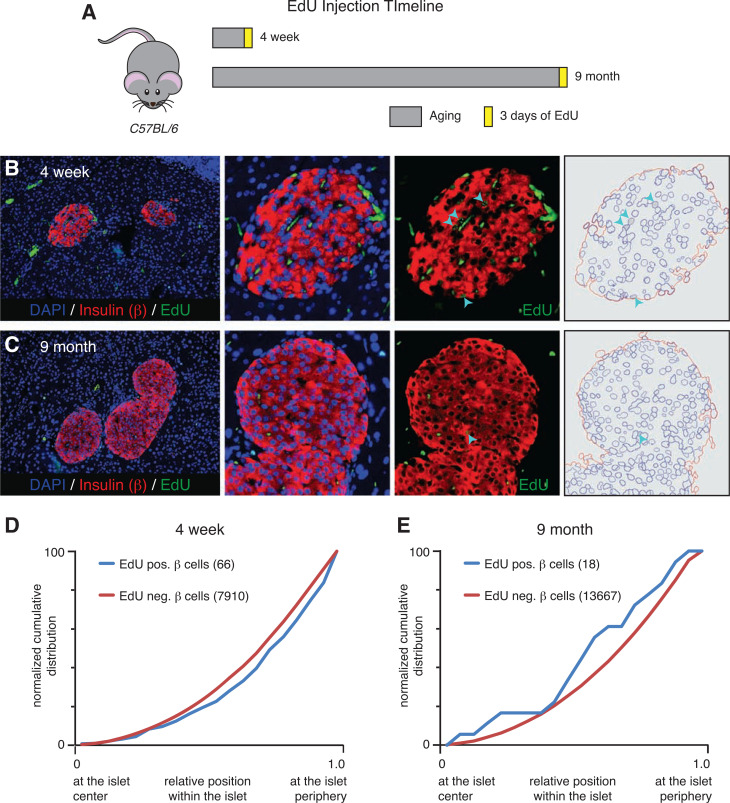
Figure 1.
Proliferating EdU-positive β-cells are randomly distributed throughout the islet. A: Schematic of EdU injection timeline of C57BL/6 mice. Representative immunofluorescence images of pancreatic tissue sections from 4-week-old (B) and 9-month-old (C) mice stained with insulin (red), EdU (green), and DAPI (blue). Arrowheads indicate examples of β-cells that are positive for EdU labeling detection. D: Normalized cumulative distribution of EdU-positive (blue) and EdU-negative (red) β-cells within 4-week-old mouse islets. E: Distribution of EdU-positive (blue) and EdU-negative (red) β-cells within 9-month-old mouse islets. For each distribution graph, n ≥ 3 mice with eight to nine islets counted per animal. neg., negative; pos., positive.

