Figure 4.
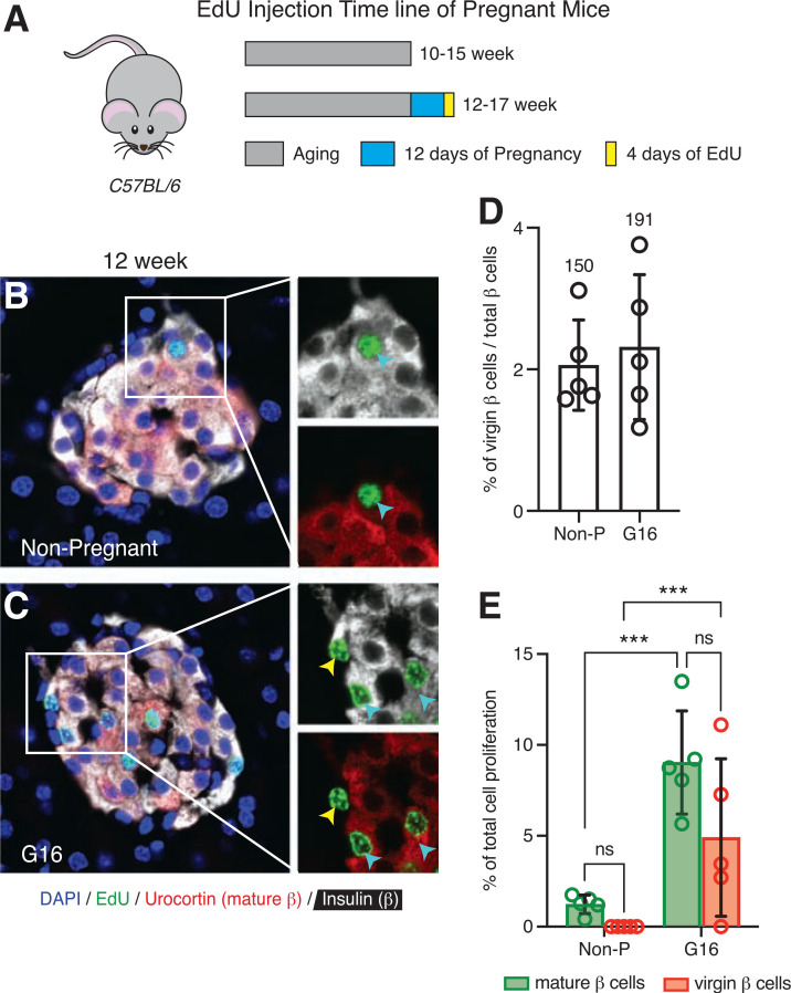
Virgin β-cell proliferation increases similarly to mature β-cell proliferation during pregnancy. A: Schematic of EdU administration timeline of pregnant C57BL/6 female mice. Representative images of islets from a 12-week-old nonpregnant (Non-P) mouse (B) and a gestational-day-16 (G16) pregnant mouse (C) immunostained with insulin (white), Ucn3 (red), EdU (green), and DAPI (blue). The cyan arrowheads point to proliferating EdU-positive mature β-cells, and the yellow arrowhead points to a proliferating EdU-positive virgin β-cell. D: Quantification of the fraction of virgin β-cells between nonpregnant and G16 pregnant mice (n = 5 per group). Values above each bar indicate the total number of virgin β-cells counted per group. E: Quantification of the total percentage of EdU-positive mature (green bars) and virgin (red bars) β-cells between nonpregnant and G16 pregnant mice over 4 days of EdU labeling period (n = 5 per group). Data were analyzed for statistical significance by two-way ANOVA, corrected for multiple comparisons with the Tukey method. Error bars represent ±SD. ***P < 0.001. ns, not significant.

