Figure 7.
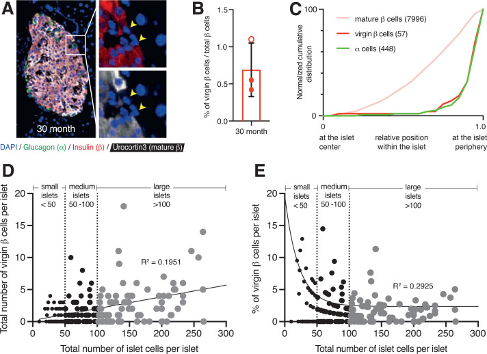
Virgin β-cells persist in 30-month-old geriatric mouse islets at their distinct location in the islet periphery and correlate positively with islet size. A: Two-dimensional representative image of a 30-month-old wild-type mouse islet immunostained with glucagon (green), insulin (red), Ucn3 (white), and DAPI (blue). The yellow arrowheads point to individual virgin β-cells that lack Ucn3 expression. B: Quantification of the fraction of virgin β-cells from 30-month-old geriatric mice. Open circles, females; closed circles, males. C: Normalized cumulative distribution of α-cells (green), virgin β-cells (red), and mature β-cells (pink) within 30-month-old mouse islets (n = 3 mice with at least 10 islets counted per animal). D: Scatter plot illustrating the distribution of total virgin β-cells in relation to islet size (measured by total β- and α-cells). Each dot represents an islet (n = 193). Relationship analyses performed using Spearman correlation and simple linear regression yielded a positive correlation (r = 0.40, R2 = 0.1951). E: Scatter plot illustrating the distribution of the percentage of virgin β-cells (measured over total β-cells) in relation to islet size. Each dot represents an islet (n = 193). Relationship analyses using Spearman correlation and nonlinear regression yielded an inverse correlation (r = −0.39; R2 = 0.2925). Islets with no virgin β-cells were excluded from this analysis.

