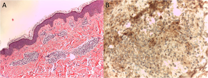
Fig. 3.
a) Histological examination showing mild hyperkeratosis and acanthosis of the epidermis; at the superficial and middle dermis, a non-specific parvocellular inflammatory infiltrate of lymphocytes, plasm cells and rare histiocytes with predominant perivasal distribution was present; dermal vessels showed thickened cell wall and endothelial cell swelling, with no clear signs of vasculitis (40x magnification); b) Immunohistochemical staining with anti-spirochetal antibodies, showing clusters of rod-shaped elements, some of which with spiral form, focally present at the epidermis and adnexal structures (100x magnification)

