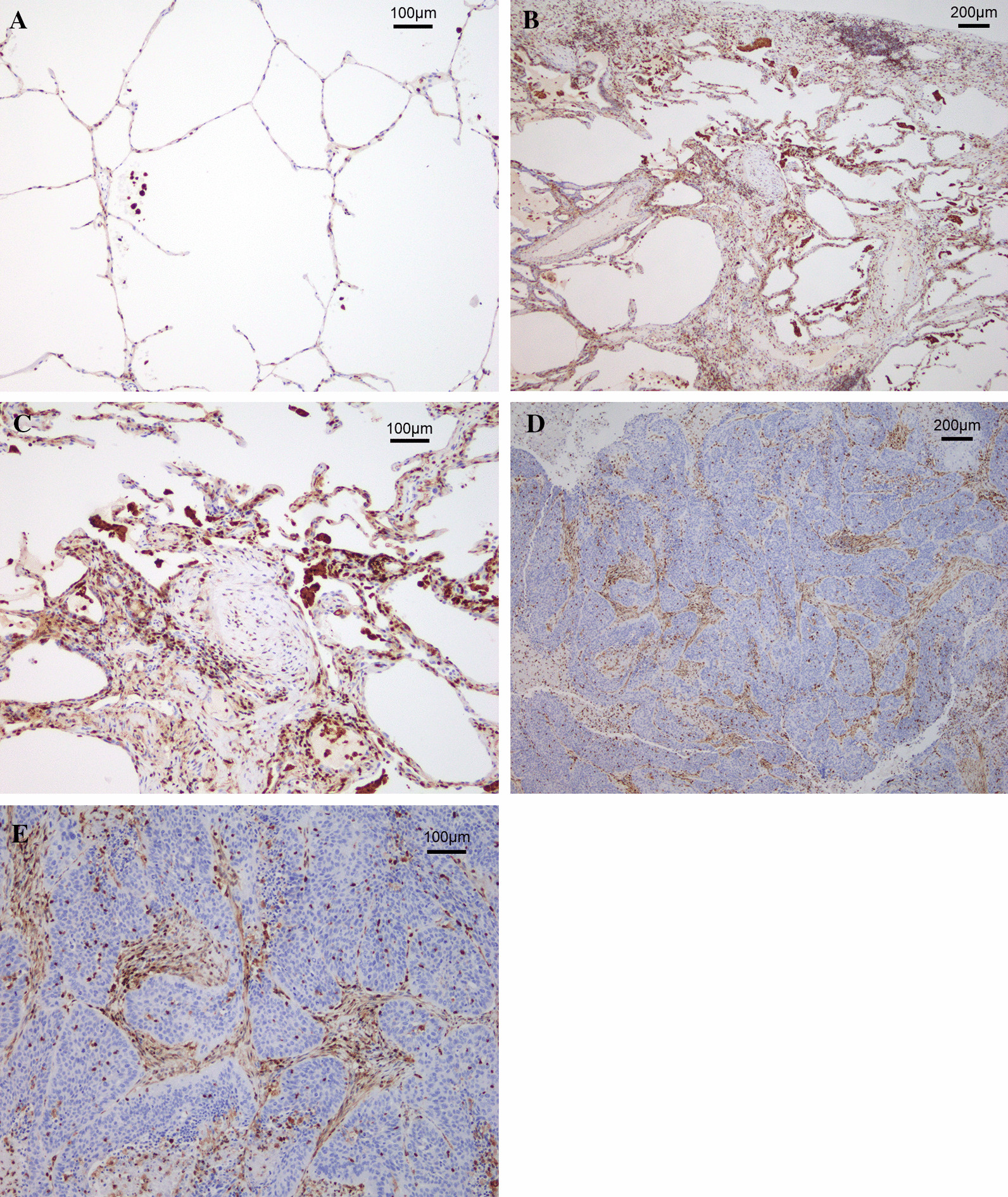
Fig. 4.

Representative images of S100A4 immunohistochemistry. A Representative image of normal lung tissue. In the normal areas of the lungs, S100A4 was sparsely expressed. B, C Representative images of areas of UIP with ×25 magnification (B) and ×100 magnification (C). The areas of UIP in all patients exhibited numerous S100A4-expressing cells such as fibroblasts, lymphocytes, and macrophages. D, E Representative images of the primary tumor with ×25 magnification (D) and ×100 magnification (E). In the primary tumor, S100A4 was expressed in the areas of stroma and fibrosis. Conversely, S100A4 was not expressed in the tumor cells
