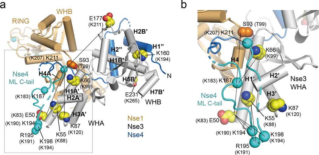
Fig. 5.
Model of the C-terminal region of XlNse4
(a) Modeling a region of the Nse4ML C-tail (residues 187 to 211) based on CL-MSNSE data. Nse1 (orange), Nse3 (white), and Nse4 (blue) are shown in cylinder representation. The modeled region is colored cyan. Solid lines indicate close spacing between Lys211 of Nse4 and Glu50, Lys87 and Glu177 of Nse3. Human Nse1, 3, and 4 residues equivalent to those of Xenopus proteins are in parenthesis.
(b) Close-up view of the Nse4ML C-tail boxed in (a). Residues 187 to 198 of the Nse4ML C-tail (cyan) are placed near the N-terminus of the H1A’ helix (Nse3, white) within ~30 Å of all crosslinked residues. Lys211 is located near Ser93 (Nse1, bright orange) and Lys66 (Nse3). Lines indicate the crosslink pairs between Nse4ML C-tail and Nse1 or Nse3. The equivalent residues in human NSE proteins are in parentheses.

