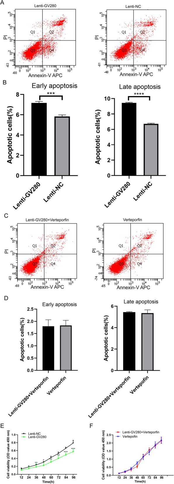
Fig. 4.

Flow cytometry analysis of apoptosis and a CCK-8 assay performed with ADS JZ SMCs. a Cells were transfected with the let-7a overexpression lentiviral vector GV280 or lentiviral null vector for 72 h and then used for flow cytometry analysis. b The percentages of apoptotic cells in the lenti-GV280 group and lenti-NC group. c Verteporfin-treated cells were transfected with the let-7a overexpression lentiviral vector GV280 for 72 h and then used for flow cytometry analysis. d The percentages of apoptotic cells in the lenti-GV280 + verteporfin group and verteporfin group. e OD450 values of the lenti-GV280 group and lenti-NC group. f OD450 values of the lenti-GV280 + verteporfin group and verteporfin group. These experiments were performed two times with three replicates in each experiment. Significance was determined by Student’s t test; ****p < 0.0001, ***p < 0.001, **p < 0.01, *p < 0.05
