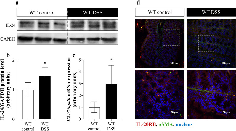
Fig. 2.
Presence of IL-24 and IL-20RB in WT mice with DSS-induced colitis. Protein level and mRNA expression of IL-24 in colon tissue (n = 6) was determined by Western blot analysis (a, b), and real-time PCR (c) by comparison with GAPDH as internal control, respectively. Results are presented as mean ± SD. *p < 0.05 vs. WT control (Mann–Whitney U-test). Localization of IL-20RB (red) in the colonic tissue samples from control and DSS-treated WT mice was determined by immunofluorescence staining with αSMA (green) co-localization (d). Nuclei are stained with Hoechst33342 (blue). Scale bars: 100 µm and 50 µm

