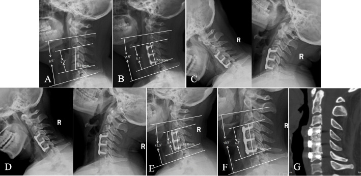
Fig. 3.
A 50-year-old female patient underwent double-level ACDF due to cervical myelopathy. A mixture of bone dust and morselized bone was used as an n-HA/PA66 cage-filling material in this patient. A Preoperative lateral radiograph of the cervical spine showing narrowing of the C4/5 and C5/6 disc spaces. B Postoperative lateral radiograph showing that the n-HA/PA66 cage increased the FSH, SSA, and C2–7 lordotic angle. C Three-month follow-up flexion-extension lateral radiograph showing the absence of motion between the spinous processes. D Six-month follow-up flexion-extension lateral radiograph showing solid fusion. E, F One-year and final follow-up lateral radiographs showing mild cage subsidence in both segments; the FSH, SSA, and C2–7 lordotic angle remained stable. G Three-dimensional CT at the 36-month follow-up time showing grade 5 fusion in both segments

