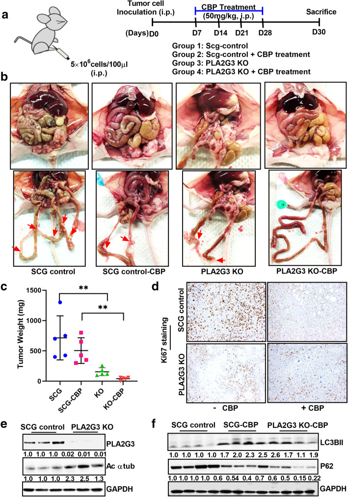
Fig. 4.
PLA2G3 KD cells are sensitized to carboplatin treatment in vivo in OC xenograft. (A) Schematic representation of the study model in mice OVCAR5 OC xenograft was provided. (B) Randomized OVCAR5 SCG-transfected control and PLA2G3 KO tumor-bearing mice were treated with or without CBP (50 mg/kg) for 4 weeks at the interval of 7 days. All mice were sacrificed on day 30. Illustrative images of the mice with the tumor burden and metastatic nodes were shown. Arrows point to the metastatic tumor nodules in the differently treated cohort of the animals. (C) Graphical presentation of the excised tumor weights in the treatment cohorts (**p < 0.01). (D) Representative images of IHC staining of Ki67 in the tissue blocks of the four treatment groups. (E) Immunoblot analysis of PLA2G3 and acetylated α-tubulin expression from the lysates of the SCG-control and PLA2G3-KO treated groups. (F) Western analysis of LC3BII and p62/SQSTM1 from the lysates of the SCG-control and CBP treated groups were shown. GAPDH is used as a loading control. Densitometric analysis using Image J software was calculated, normalized to GAPDH, and fold change was provided beneath the panel in both cases

